The relationship between phycobiliprotein, phycocyanin and phycobilin
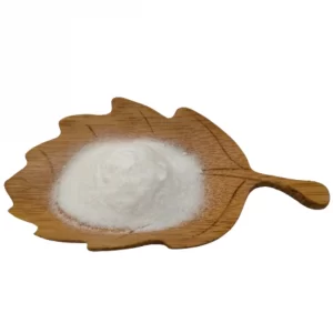
Phycocyanin is also known as phycocyanin, phycocyanin, and cyanobactin; phycocyanin is a type of phycobiliprotein.
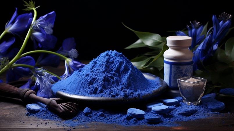
Phycobiliprotein mainly exists in cyanobacteria, red algae, cryptophytes and a few dinoflagellates. Its main function is to serve as a light-harvesting pigment complex for photosynthesis. In some algae, phycobiliprotein can also be used as a storage protein to enable Algae survive during seasons when nitrogen sources are scarce. Known phycobiliproteins can be mainly divided into four categories, namely Phycoerythrin, Phycocyanin, Phcoerycyanin and Allophycocyanin. According to different sources, each major category can be divided into several subcategories. Each subcategory is preceded by B, C, and R. These letters are used to indicate the source of phycobiliprotein. If classified completely according to spectral type, phycobiliproteins can be divided into 6 categories, namely: allophycocyanin B (APB), allophycocyanin (AP), phycocyanin (PC), phycoerythrocyanin (PEC), phycoerythrin I (PEI) and phycourobilin-containing phycoerythrin II (PEII) in cyanobacteria. Phycocyanin, or phycocyanin, is an indicator pigment for cyanobacteria in water, and its concentration reflects the biomass of cyanobacteria.
Phycobilin, the chromophore of phycobiliprotein, is an important photosynthetic pigment that absorbs light energy and transmits excitation energy. In 1928, Lemberg first demonstrated that phycobiliprotein is composed of an apoprotein and a chromophore with a tetrapyrrole structure. O’hEocha acidolyzed phycobiliprotein with 12N hydrochloric acid in 1958 to obtain the chromophore. In 1966, he used methanol reflux to separate the chromophore. Schram and Kroes also used HBr to treat phycobiliprotein in 1971 and obtained the free chromophore. , they called it “Phycobilin”. And they found that C-phycocyanin only contains one chromophore, namely Phycocyanobilin (PCB). In 1964, Ocarra discovered that in addition to phycoerythrobilin in R-phycoerythrin, he also discovered another phycobilirin. Its absorption peak is located at 495nm. It is difficult to remove it from the detachment when treated with concentrated hydrochloric acid at room temperature. After being removed from the base protein, this chromophore was later named “Phycourobilin (PUB)”. The structures of the above-mentioned color bases were successively determined. Bryant et al. discovered the fourth chromophore “Phycobiliviolin” from the α subunit of phycoerythrocyanin in 1976. Its structure was determined by Bishop et al. in 1987. The only species of phycobilins known so far are phycoerythrobilin (PEB), phycocyanobilin (PCB), phycourobilin (PUB) and phycobiliviolin (PXB). ), their basic chemical structures are the same. In different phycobiliproteins, the type and content of phycobilin are different. These four types of phycobilins are isomers, and their differences are reflected in the different positions of the double bonds.
Phycocyanin supplier: www.backvita.com
Email: [email protected]
Phone: +86 (029) 8187 2325